How Bjørn Angelsen’s innovation spearheaded a medical ultrasound industry
The story of Norway’s medical ultrasound industry starts in the early 1970s in Trondheim when a young doctor, Alf Brubakk, who was working at the regional hospital (now St Olav’s Hospital) met an ambitious engineering student, Rune Aaslid, from the Norwegian Institute of Technology (now the Norwegian University of Science and Technology (NTNU)), who claimed he could make a mathematical model of the cardiovascular system. Together, they created Jenny, a patient simulator built using analogue electronics and named after the first real patient their device was used on [1, 2, 3].
As they worked on their model, it became clear to Aaslid and Brubakk that the theory on blood-flow simulations required experimental data. Could ultrasound be a useful means of acquiring such data?
The solution came with Bjørn Angelsen: compelled by the mathematical challenges of the project combined with his intimate knowledge of ultrasound waves, he developed a device called the Pulsed Echo Doppler Flowmeter (PEDOF) that could accurately measure blood flow velocities in the aorta and heart based on the Doppler effect.
Pioneering the “E = mc2 of cardiology”
The 1960s and 70s saw an explosion of new possibilities in cardiac surgery thanks to technological developments and safer anaesthesia.
“Cardiac surgery was the real challenge at that time, everyone in the medical field was focused on that. The cardiologists were focused on invasive techniques to measure pressures in the heart, which means putting in catheters. And so, at the time ultrasound looked a little sort of esoteric,” Angelsen reminisces. “But then along came a doctor who had a very interesting background, and he completely changed this perspective.”
The doctor Angelsen refers to was Jarle Holen, who had previously worked as an aerodynamics engineer on the Boeing supersonic jet project in Seattle in the 1960s. In collaboration with Rune Aaslid, he used his aviation background and knowledge of fluid dynamics to apply Bernoulli’s principle to heart valves. Together, they calculated pressure gradients in patients with narrowed heart valves using estimates of blood flow velocities from the PEDOF device [4]. This made it possible to calculate the pressure inside the heart using harmless ultrasound waves, thus avoiding invasive catheterisations.
Building on this innovation, Angelsen experimented on optimal sensitivity Doppler equipment that could be used in pulsed wave (PW) to obtain spatial resolution inside the heart cavities and in continuous wave (CW) to measure the high velocities that occur in heart valve lesions. Another challenging task he tackled was real-time estimation of the Doppler frequency to give time-variable curves of the blood velocities inside the beating heart.
Angelsen further simplified and introduced material parameters into the Bernoulli equation, resulting in the formula P= 4V2, where V is the velocity and P the pressure gradient. This simple yet powerful formula rapidly gained acceptance among clinicians and was later nicknamed the “E = mc2 equation of cardiology”.
“I’ve always loved mathematical theory. When you’re too focused on commercial strategy and outselling your competitors, you risk losing your edge; without developing new theory it can be hard to keep track and to move the field forward.”
Bjørn Angelsen
Professor
An ultrasound imaging unit fit for industry
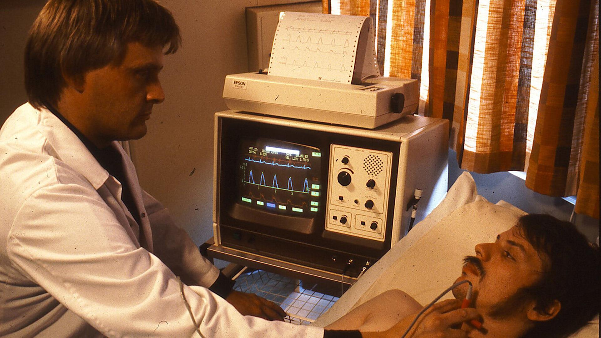
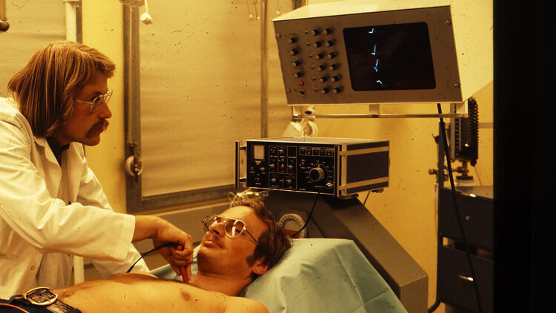
The next step was to establish clinical trials utilising the new technology. In summer 1976, Angelsen employed five engineering students at NTNU to assemble 10 Doppler ultrasound units. The team sold four of the devices, and the rest stayed in Trondheim.
In those early days, the instrument did not have an imaging unit and was difficult to use in the clinical setting. Despite these challenges, cardiologist Dr Liv Hatle used one to pioneer a new technique for diagnosing heart disease (read the article here), while Dr Sturla Eik-Nes used another to study foetal blood flow in pregnant women [5].
To improve clinical usability, the engineers made many upgrades, taking advantage of the analogue-to-digital electronics boom happening at the time. Angelsen emphasises that “the work we did came in parallel with this development of instrumentation, integrated circuits and computers.”
The researchers and clinicians in Angelsen’s group in Trondheim knew that they needed an industrial partner to develop an imaging unit for the PEDOF technology and to launch it commercially, so they turned to a small start-up based in Horten called Vingmed AS.
Together they created the CFM 700, the world’s first annular array colour flow imaging system. The system was introduced to the world at the American College of Cardiology meeting held in Atlanta, Georgia in March 1986.
“Before going to the conference, we tested the device on ourselves,” explains Angelsen. “By chance we came across a patient in the hospital hallway who had some strange chest pain, so we used the device on him and diagnosed a critical aortic condition. This patient was admitted to surgery immediately and was saved! Very different regulatory requirements from what inventors must adhere to today.”
The demo at the conference was eventful, Angelsen recounts. “We had to transport the device to the top floor of the building for a demonstration, but as soon as we stepped out of the elevator it stopped working. The screen was full of strange signals – we couldn’t understand it. Then we looked out of the window and noticed a big antenna system across the street whose signals were interfering with our device. Fortunately, we moved down to the cellar and it worked perfectly.”
The CFM 700 went on to become an immediate commercial success, selling a total of 4 500 units.
Inspiring a new generation of healthcare innovators
Four of Angelsen’s students who had worked on the first Doppler prototypes in summer 1976 later became important figures in Norwegian tech. Kjell Kristoffersen became Head of Technology at GE Ultrasound, Arne Grip founded Medistim, Sverre Horntvedt became CEO of Sensonor, and Veroslav Sedlak became Head of Business Development at Goodtech.
Undoubtedly, Angelsen’s invention has laid the technological and theoretical basis for a thriving Norwegian medical ultrasound industry. GE Healthcare has incorporated much of this innovation into its ultrasound equipment and services market, which now reports annual revenues of USD 3 billion [9].
Despite a monumental career in medical ultrasound spanning 50 years, Bjørn Angelsen has never rested on his laurels and shows no signs of slowing down. In 2010 he founded the start-up SURF Technology as a spin-off from his research group at NTNU and continues to inspire the next generation of healthcare innovators.
Spotlight on Norwegian ultrasound industry
This article was written by Leah Anderson on behalf of EXACT-Therapeutics AS, Innovation Norway, Norway Health Tech and Investinor AS. The article is part of the series “The global reach of Norwegian ultrasound innovation”, which focuses on how Norwegian ultrasound innovation is impacting medicine globally.
